
Figure 16: Chemical structures of the prototypical linear furocoumarin (psoralen), the prototypical angular furocoumarin (angelicin), and the two most clinically relevant photosensitizing psoralens (8-methoxypsoralen and 5-methoxypsoralen). 15
Phytophotodermatitis connotes phototoxic reactions consisting of erythema (with or without blistering) and delayed hyperpigmentation. Phototoxicity implies an immediate or delayed inflammatory reaction caused by the combination of a topical or oral photosensitizing agent followed by the appropriate wavelength of ultraviolet radiation (UVR) to the skin.37 This response can occur in any person who has been exposed to adequate amounts of a photosensitizing chemical and UVR. It is therefore not an immunologic reaction, and no prior sensitizing exposure is necessary for any potential victim.
HISTORY
Plants have been known to cause hyperpigmentation since antiquity. As early as 2000 B.C. in Egypt, the juice of Ammi majus (Umbelliferae), false Bishop's Weed, that grows throughout the Nile River valley as a weed, was rubbed on patches of vitiligo after which patients were encouraged to lie in the sun. Even today, Egyptian herbalists sell Aatrillal, a yellowish-brown powder made from Ammi majus seeds for the treatment of leukoderma. Indian medicine used boiled extracts of leaves, seeds, and roots of Psoralea corylifolia (Leguminosae) a South African native known as the bavachee. Topical or systemic figs followed by sun exposure was also used to treat 'white leprosy,' as vitiligo is still called in India. Chinese herbal medicine even today recommends that leaves of Angelica archangelicum (Umbelliferae) be used. Our name for the chemical family of phototoxic ingredients, psoralens, was derived from Psoralea corylifolia.1,38
In 1897, reports of dermatitis following contact with parsnips, Pastinaca sativa, (Umbelliferae) and/or angelica, Angelica archangelicum, came from both the United States and England.39, 40 Unfortunately, neither author recognized the necessity of UVR for the reaction. In 1916, Freund observed characteristic hyperpigmented lesions resembling pendants that he attributed to eau de cologne containing bergamot oil. 41 He, too, did not recognize the necessity of UVR for the cutaneous changes. In 1932, Oppenheim reported that the combination of meadow grass exposure and UVR could result in a striate arrangement of itching blisters 24-48 hours later.42 In 1938, Kuske showed that plant furocoumarins caused photosensitization.43 In 1939, Jensen and Hansen reported that UVR wavelengths between 320 and 380 nm (UVA) caused the maximal reactions.44 Klaber introduced the term 'phytophotodermatitis' in 1942 to emphasize the necessity of plants and light to cause the reaction.45
It was an Egyptian, Professor Abdel Monem El Mofty, Department of Dermatology, Cairo University Medical School, who observed plants used in Egyptian folk medicine and began the development of modern photochemotherapy (PUVA) for vitiligo and psoriasis. In the 1940s, he used crystalline methoxsalen (8-MOP, xanthotoxin) followed by sunlight exposure to treat vitiligo. The Department of Dermatology at Harvard Medical School first successfully treated psoriasis with PUVA in 1974. Today, PUVA is used in the therapy of many other diseases including palmoplantar pustulosis, mycosis fungoides, atopic dermatitis, and generalized lichen planus.38
FUROCOUMARINS
Furocoumarins are likely related to a plant's natural defense against fungal attack. Apium graveolens, fresh celery, contains 10-100mg/g wet weight of psoralens in healthy plants, but may have 320 mg/g when infected with Sclerotina sclerotium (pink rot disease). Disease-resistant celery unfortunately possesses high levels of furocoumarins concentrated in the leaf sap and stem sap. Only 1mg of 8-MOP per square centimeter of skin is necessary to produce blisters after 2.4 J/cm2 (less than 10 minutes of summer sunlight in Colorado)! In one study, 163/320 (51%) randomly selected Michigan celery workers displayed vesicular and bullous dermatitis on the fingers, hands, and forearms. In this study, the authors could only induce phytophotodermatitis with celery infected with pink-rot disease. Furocoumarins are tricyclic compounds produced by the fusion of a furan ring (positions 1' to 5') with a bicyclic benz a-pyrone to form both linear (psoralen) and angular (angelicin or isopsoralen) structures (Figure 4). 1, 15 ,46
|
|
|
Figure 16: Chemical structures of the prototypical linear furocoumarin (psoralen), the prototypical angular furocoumarin (angelicin), and the two most clinically relevant photosensitizing psoralens (8-methoxypsoralen and 5-methoxypsoralen). 15 |
Two forms of furano-coumarin condensation occur in nature. The first forms linear molecules by linking the 3'2' furan bond to the 6,7 coumarin bond. The 3'2' bond may also link to the 7,8 coumarin bond to produce angular furocoumarins. In general, the linear furocoumarins (psoralens) are more phototoxic than the angular furocoumarins (angelicins). The only exception to this is the angular furocoumarin known as pimpinellin found in members of the genus Heracleum (Umbelliferae). The most severe reactions occur with 5-methoxypsoralen (5-MOP; bergapten) isolated from Citrus bergamia (Rutaceae), the bergamot orange, and 8-MOP (xanthotoxin) isolated from Fagara xanthoxyloides (Rutaceae). Psoralen is more phototoxic than 8-MOP and 5-MOP.1, 15
MECHANISMS OF PHOTOTOXICITY 1, 15 , 46
The phototoxic effect of furocoumarins relies on their ability to absorb photons. After forming short-lived, high energy states, the energy is released and causes subsequent cellular damage.46
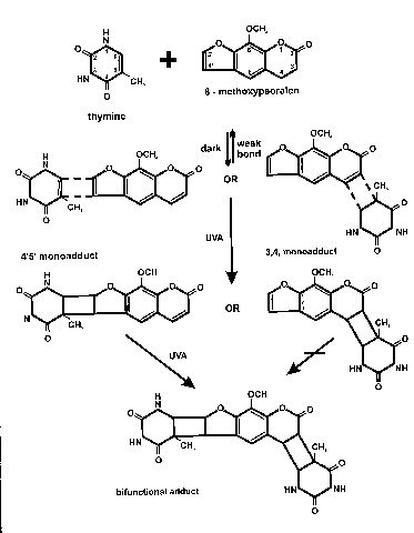
Two distinct but concurrent reactions take place when psoralen-treated skin is exposed to UVA radiation. Psoralens have an absorption peak at 300 nm and an action spectrum peak at 335 nm.46 In an oxygen-independent reaction, UVA excites psoralens to a triplet state that causes covalent binding of the psoralen molecule with nuclear DNA. Monofunctional adducts form between the 4'5' furan bond or the 3,4 coumarin double bond and the 5,6 bond of a pyrimidine (cytosine or thymidine). When the monoadduct is formed by the 4'5' furan bond, that product may form bifunctional, interstrand cross-links between pyrimidine bases. (Monoadducts of a pyrimidine and the 3,4 coumarin double bond do not go on to form bifunctional adducts.)
|
|
|
Figure 17: 8-MOP photoadduct formation with thymine molecule |
Although monoadducts may produce mutations, inhibit DNA synthesis and cell proliferation, stimulate melanogenesis, and cause cell death, these effects are greater with cross-link formation. The only monoadduct formation that can cause significant phototoxic bullae is that formed by pimpinellin, an angular furocoumarin found in Heracleum spp. Only the linear psoralens can form bifunctional, interstrand cross-links, and these cross-links to keratinocyte DNA are the main chemical change responsible for severe skin damage after UVA.
While oxygen-independent psoralen reactions cause the most cellular damage, oxygen-dependent (photodynamic) reactions also cause clinical effects. Reactive oxygen species form by the interaction of psoralens and oxygen. These occur in cell nuclei, in cell membranes of epidermal, dermal, and endothelial cells, and in cytoplasmic constituents such as enzymes, RNA, and lysosomes.
Psoralen-induced hyperpigmentation occurs via a multitude of interrelated changes. After bifunctional photoadducts are formed in keratinocyte and melanocyte DNA, increased mitosis of basal layer keratinocytes and melanocytes is observed. In fact, the melanocyte population will double or triple within 3-7 days. Melanocyte hypertrophy and increased arborization is also seen. Tyrosinase activity increases to make more melanin that is packaged into an increased number of melanosomes found in melanocytes and transferred to malpighian layer keratinocytes. In fair-skinned individuals, the distribution of melanosomes changes. Instead of small, clumped melanosomes in keratinocytes, large, single, dispersed melanosomes are seen capping the nuclei. This pigmentation can last for months or years.
Non-psoralen photosensitizing chemicals have been isolated from members of the daisy family (Compositae): polyacetylenes (such as phenylheptatriene) and thiophenes (such as a-terthienyl). However, phytophotodermatitis after contact with members of the Compositae have never been reported. Therefore, it is likely that these phototoxic compounds do not contact the skin in high enough concentration or that they do not penetrate the stratum corneum.
CLINICAL ASPECTS OF PHYTOPHOTODERMATITIS
Because phytophotodermatitis is non-immunologic, it may be elicited in anybody given sufficient furocoumarins and UVA. Phototoxic reactions can occur from UVR exposure only 15 minutes after contact with topical furocoumarins, but UVA sensitivity peaks 30-120 minutes after contact.46
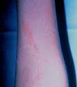
Bizarre configurations of erythema, edema, and bullae appear after a latent period of roughly 24 hours. The inflammatory reaction peaks at 72 hours. These painful, non-pruritic reactions are more often seen in middle to late summer when psoralen concentrations are highest in the offending plants and more skin is exposed to direct sunlight. Wet skin, sweating, and heat enhance the phototoxic response.1 To avoid misdiagnosis as poison ivy dermatitis, it should be noted that the initial erythematous and bullous reaction only occurs in sun-exposed areas. Hyperpigmentation follows one to two weeks after UVR exposure and lasts months to years. Interestingly, areas affected by phototoxic reactions may remain hypersensitive to UV light for years.15
Erythematous vesicles Histology slide of skin with severe sunburn
Figure 18: Clinical and Pathological
Characteristics of Hypersensitivity recation.

Patients can be exposed to furocoumarins in many settings (Table 4). Modern power tools such as the 'Weed-Eater' or 'Strimmer' (string-trimmer) delivers a buckshot spray of weeds including many Umbelliferae. 'Strimmer Dermatitis' appears 12-24 hours later as red irregular macules and papules, not bizarre linear and angular streaks, on the anterior chest and arms. The most commonly implicated species are members of Umbelliferae: the cow parsnip (Heracleum sphondylium), giant hogweed (Heracleum mantegazzianum) and cow parsley (Anthriscus sylvestris). 47, 48
Berlock dermatitis comes from the German word 'berlock' or the French word 'breloque' meaning a trinket or charm. Rosenthal coined the term in 1925 to describe pendant-like streaks of pigmentation on the neck, face, arms, or trunk. He suspected that they were due to drops of fluid. He did not know that, in 1916, Freund had described pigmented macules due to sun exposure after the application of cologne water.41 The phototoxic ingredient was bergamot oil found in the fresh rind of Citrus bergamia, a small orange. Mainly grown in southern France and Italy, bergamot oranges are processed into crude bergamot containing 0.3-0.4% 5-MOP ('bergapten' from 'bergamot'). Still today, natural bergamot oil is used in some parts of the world in suntanning creams and in certain brands of tea.46 Bergamot oil has been applied to the upper lips of women in theater since its irritancy could induce a 'pouty' lip when necessary. Since the introduction of artificial oil of bergamot, 'berlock' dermatitis has become rarer.1, 15
Figure 19: Berloque
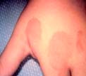
Phytophotodermatitis has been mistaken for child abuse. A 13-month-old girl presented with red finger marks on both shoulders. Although not typical of bruising, initial medical evaluation was that of child abuse. History revealed that the mother had been gardening that day and had cut down an Umbelliferae family member, probably giant Russian hogweed (Heracleum mantegazzianum).22 Two other children were noted to have irregular digitate hyperpigmentation on their backs, sometimes simulating hand print configuration. They had been on beach vacations in Mexico with their parents. While squeezing limes to make drinks, the parents frequently touched their children's exposed skin. Multiple hues were not seen in the skin lesions as would be expected in healing bruises.49
|
|
|
Figure 20: Lime phytophotodermatitis and late hyperpigmentation |
|
Gardening |
Brushing against Dictamnus spp. (USA, Europe, N. China) or Ruta (UK) Cultivating celery, parsnip, or parsley Clearing weeds with a 'weed wacker' (USA) or 'string trimmer' (UK) Pruning or harvesting figs Growing Angelica for herbal medicine (Korea), cake decorating (when candied), tonic |
|
Canning or Processing fruits and vegetables |
Canning celery or stocking celery in grocery stores50 Making lemonade or limeade, especially if selling it outside51,52 Squeezing lime juice for margaritas and other drinks53 |
|
Hiking/Jogging/Walking |
Through fields and riverbanks (Heracleum spp.) (Pacific Northwest, Europe) Rolling in the meadow Hiking in southern California and Baja California, Mexico (Cneoridium dumosum) - Coast spice bush (Rutaceae)54 |
|
Medications and Cosmetics |
Application of 'tan promoters' or perfumes containing bergamot oil (berlock dermatitis) Excessive UV exposure after taking or applying psoralens for PUVA Application of rue (Ruta spp.) as an insect repellant |
|
Play |
Making peashooters with Heracleum mantegazzianum (giant Russian hogweed) or other Heracleum spp. Playing amongst rue bushes and Umbelliferae Fighting with parsnips/celery |
|
Ingestion |
Ingestion of excessive psoralens (esp. celery) before using a tanning parlor (UVA) Ingestion of Chlorella (Japan) |
|
Clothing |
Wearing leis of Pelea anisata (Hawaii)55 Fig Leaf |
Limes have gained a reputation as potent photosensitizers causing their own 'Lime Disease'.51 They contain psoralen, 8-MOP, and 5-MOP which possess relative phototoxic activities of 100, 37.5 and 27.5 respectively. The most effective photosensitizer in limes (because of its concentration) is bergapten (5-MOP) which is 13-182 times more plentiful in the rind than the pulp. Arizona Persian limes contain six times as much 5-MOP as Florida Key limes.52 As little bergapten as 10mg/g will suffice to allow a phytophotodermatitis. While pulp concentrations are about 1 mg/g in limes, rind concentrations are 20-130 mg/g.52 Non-dermatologists frequently misdiagnose phytophotodermatitis due to lemons and limes. In ten successive cases referred to dermatologists in San Diego County, CA, they were variably diagnosed as allergic contact dermatitis from poison oak, atypical bruising: rule out malignancy, cellulitis, child abuse, erythema multiforme, fungal infection, impetigo, infectious lymphangitis, and thrombocytopenic purpura.51
Figure 21: Phytophotodermatitis
from sucking lemons

Phototoxicity following ingestion of naturally-occurring psoralens appears to be rare. In 1986, Pathak reported a study of two fair-skinned 'volunteers' who ate 20 stalks of celery, 25 dried figs, and 250 g of parsley. After exposure to midday sun for 30 minutes, no phototoxic reaction occurred.1 Oral ingestion of large quantities (450g) of celery (equivalent to 45 mg of pure psoralens) followed by time in a UVA tanning bed one hour later led to widespread phytophotodermatitis.56 Celery broth (250 cc) ingested two hours before PUVA therapy for widespread lichen planus caused severe, widespread phototoxicity.57 The concentrations of psoralens are high enough in some vegetables (4 mg/100 g parsnip root and 5-32 mg/100 g celery root) to be possible photosensitizers when ingested in large quantities.57
PHOTOTOXIC PLANTS
Four main plant families have been implicated as causing phytophotodermatitis: Umbelliferae (Apiaceae), Leguminosae (Fabaceae), Moraceae, and Rutaceae.
Bergapten (5-MOP), followed by xanthotoxin (8-MOP), are the most commonly found linear furocoumarins in phototoxic plants. Psoralen is uncommonly found or is found at low concentrations. The angular furocoumarins such as pimpinellin, angelicin, and sphondin have a much smaller contributing effect to phytophotodermatitis.
Umbelliferae (Apiaceae)
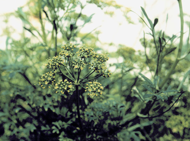
Umbelliferae is the main family of plants implicated as causing phytophotodermatitis. This family of plants is very easy to identify in the wild because of their distinctive floral structure. Numerous small flowers are held in an 'umbel' (a cluster of flowers on stalks of roughly equal length arising from a single point).7
 |
 |
|
|
|
Often, the flower heads are made up of many small umbels to make 'compound umbels (Figure 22).' When fruits are present, they are small, oblong or cylindrical. The flower heads are sheathed at the base by one or more leaf-like bracts. The various family members look so similar, that precise identification of species can be confusing.
Ammi majus is the world's major source of xanthotoxin (8-MOP). It has been found naturally in Egypt's Nile valley, Europe, India, Russia, the United States, and South America.1 Furocoumarins are especially abundant in roots and leaves of the Umbelliferae.
|
|
|
|
Figure 23: Ammi majus, Queen Anne's Lace, compound umbel |
Ammi majus, fruit |
The roots of Angelica gigas Nakai (Korean angelica) are used in Korean herbal medicine as a diuretic and to treat anemia and hemorrhoids. Working in angelica gardens has caused gardeners to develop phytophotodermatitis. Photopatch testing revealed that the fruit and seeds of the plant, but not the leaf and stem, contain sufficient furocoumarins to cause phototoxic reactions.47
Table 6. Important members of the Umbelliferae implicated in phototoxic reactions
|
|
|
|
|
Ammi majus |
false Bishop's weed |
Historical and economic importance: 2000 BC vitiligo treatment. Major source of 8-MOP. |
|
Angelica archangelica |
Angelica |
Candied portions used in cakes Characteristic Benedictine flavor |
|
Angelica sylvestris |
Wild angelica |
|
|
Anthriscus sylvestris |
Cow parsley, wild chervil |
'Strimmer dermatitis' |
|
Apium graveolens |
Celery |
Infected plants have more psoralens |
|
Carrot |
|
|
|
Foeniculum vulgare |
Fennel |
|
|
Heracleum laciniatum |
Tromsø Palm |
Scandinavia |
|
Heracleum lanatum |
Cow parsnip |
North America |
|
Heracleum mantegazzianum |
Giant Russian hogweed, wild rhubarb, hogweed tree |
Giant weed naturalized across UK, Canada, USA |
|
Heracleum sphondylium |
European cow parsnip, hogweed, cow parsley |
Main cause of 'strimmer dermatitis' |
|
Pastinaca sativa |
Parsnip |
Furocoumarins in leaves, roots, stem, fruits |
|
Parsley |
|
Heracleum sphondylium, cow parsley, is thought by some to be the major cause of phytophotodermatitis in Europe and North America.1 Once introduced into a new area, Heracleum spp. tend to grow out of control up to 10 feet tall. The black seeds and leaf extract contain the most potent photosensitizers.1 The greatest threat from Heracleum occur in the autumn when the weather favors the development of many seeds.46 Hollow stems of H. mantegazzianum and H. laciniatum (Tromsø palm, Norway) used as peashooters and trumpets have caused perioral blisters in children.15 Flowers, fruit, leaves, and roots of H. laciniatum are much more phototoxic than the stems.58 Heracleum spp. contain both 5-MOP and 8-MOP.
|
|
|
|
Figure 24: Hercaleum mantegazzianum, hogweed |
H.mantegazzianum (compound umbel) |
The concentration of furocoumarins may increase in response to fungal attack. Apium graveolens, fresh celery, contains 10-100mg/g wet weight of psoralens (5-MOP, 8-MOP, and 4,5',8-trimethylpsoralen) in healthy plants, but may have 320 mg/g in specimens infected with the fungus Sclerotina sclerotium (pink rot disease). Psoralens appear to be 'phytoalexins,' substances produced by the plant to resist further fungal attack. Disease-resistant celery has been developed, but these have increased levels of natural furocoumarins.15
Celery harvesters and canners are at high risk for developing phytophotodermatitis. Only 1mg of 8-MOP per square centimeter of skin is necessary to produce blisters after 2.4 J/cm2 (less than 10 minutes of summer sunlight in Colorado)!15 In one study of 320 randomly selected Michigan celery workers, 163 (51%!) displayed various stages of vesicular and bullous dermatitis on the fingers, hands, and forearms.59 In this study, the authors could not induce phytophotodermatitis except with celery infected with pink-rot disease (Sclerotinia sclerotiorum). The furocoumarins are often concentrated in the leaf sap and stem sap.
|
|
|
Figure 25: Apium graveolens, celery |
Less than 0.1 g linear furocoumarins/100 g dried plant can elicit phototoxic reactions. The following table15,46 shows the concentrations of 5-MOP and 8-MOP in various Umbelliferae members.
(g/100 g dried plant) (g/100 g dried plant) Ammi majus Pastinaca sativa Heracleum laciniatum H. nipponicum Apium graveolens (infected)
Rutaceae
The Rutaceae family includes tropical (Citrus spp.), subtropical, and temperate (Ruta spp.) climate plants and is the second most widely distributed family of plants reported to cause phytophotodermatitis.1 Many Rutaceae grow as shrubs or small trees with flowers having four or five sepals and petals and fleshy fruit. A major cause of phototoxic reactions in the United States, especially in Florida and the desert southwest, is the rind of the Persian lime, Citrus aurantifolia. Bartenders commonly develop vesicles on the first and second fingers from squeezing limes to make cocktails.1 Phototoxic reactions were also reported among day camp children in Maryland using Persian limes to make sweet-smelling pomander balls.60 While limes are notorious for this, even the sweet orange, Citrus sinensis, can cause a phototoxic cheilitis after contact with the rind.61 Norwegian skiers often suck sweet oranges while skiing in the mountains. They sometimes return home with a lower lip cheilitis. However, the essential citrus oils in sweet oranges may also cause a chemical irritant dermatitis.61
Figure 26: Citrus medica,
citron

Ruta graveolens, garden rue, is a native sub-shrub of the Mediterranean area with a long history as a folk-medicine remedy for urticaria, warts, and erysipelas. Its taste and small is incredibly bitter. It is probably the most common cause of phototoxicity acquired in English gardens. This plant contains 5-MOP (bergapten), 8-MOP (xanthotoxin), and angelicins.15
Figure 27: Ruta graveolens,
garden rue

Dictamnus albus, the gas plant or burning bush, may have been the burning bush encountered by Moses on Mount Sinai.15 The aromatic oil exuding from the plant can be ignited briefly without harming the plant.46 It has become a common yard plant in the United States and Canada, and it grows wild in central and southern Europe, eastern Siberia, and northern China.15,46 The seed pods have high concentrations of both 5-MOP and 8-MOP.15
The coast spice bush, or berryrue (Cneoridium dumosum), is another member of the family Rutaceae recently implicated as a cause of phytophotodermatitis.54 This bush grows on mesas and bluffs below 2,500 feet in the chaparral (sage brush) vegetation zone of southern California and Baja California, Mexico. C. dumosum is a three to six foot tall, slender-twigged, highly-branched evergreen shrub. More than 20 students on a field trip to Mexico acquired phototoxic dermatitis after contact with the plant.54
Reactions to lei flowers worn around the neck, specifically Pelea anisata (Rutaceae), the mokihana, have been reported in Hawaii. These flowers are picked from mountain forests on the island of Kauai during the fruiting season. They release essential oils with pleasant, anise-like (licorice) aromas. These reports are the greatest after King Kamehameha day in mid-June.55
|
Citrus aurantifolia |
Lime |
|
Citrus aurantium |
Bitter orange |
|
Citrus bergamia |
Bergamot orange |
|
Citrus limetta |
Sweet lemon |
|
Citrus limon |
Lemon |
|
Citrus paradisii |
Grapefruit |
|
Citrus sinensis |
Sweet orange |
|
Dictamnus albus |
Burning bush, gas plant |
|
Phebalium argentuem |
Blister plant |
|
Pelea anisata |
Mokihana |
|
Ruta graveolens |
Rue |
Moraceae
The last family of phototoxic importance is the mulberry family. Ficus carica (the fig tree) is native to the Middle East and has been widely cultivated throughout warm, temperate regions of the world. No other food source is mentioned more in the Bible, and the milky juice has been used to destroy warts and cure skin infections. In A.D. 50, Dioscorides noted that vitiligo would repigment if 'cataplasmed with ye leaves or ye boughes of ye Black Figge.'26
Figure 28: Ficus carica,
fig Figure 29: Ficus aspera,
immature figs


Psoralen and bergapten (5-MOP) are found chiefly in the sap of leaves and shoots.62 Surprisingly, and unlike Ammi majus, no furocoumarins were detected in ripe or unripe fruit. Some studies have found low concentrations of furocoumarins in the milky sap of green fruit stalks, but others have not.62 Furocoumarin levels in figs are higher in April and July than in October.62
Leguminosae (Fabaceae)
The main source of psoralens for treatment of vitiligo in India is Psoralea corylifolia, the bavachee or scurf-pea. The genus lent its name to its phototoxic chemical constituents. It has been used to treat vitiligo since 1400 B.C., and its seeds are still used for this purpose.26 This genus also includes Myroxylon balsamum and Myroxylon pereirae from which balsam of Peru is extracted. 9, 15
Figure 30: Psoralea
acaulis

Other Families
Compositae (daisy family), Ranunculaceae (buttercup family), Brassicaceae, Convolvulaceae, Hypericaceae, and Anacardiaceae have been reported to contain furocoumarins and other photosensitizing chemicals, but none of these has been shown to cause clinical phytophotodermatitis.1
Phytophotoallergic contact dermatitis
According to Lovell,15 no true photoallergic reaction to plants has been proven except perhaps one case from contact with Parthenium hysterophorus (Compositae).63 However, Kavli et al. reported the induction of photoallergic contact dermatitis to Heracleum laciniatum.64 Lovell comments that the sensitizing agents in this case were poorly phototoxic constituents such as isobergapten and sphondin.23 Pathak has stated that the contact photoallergic reactions reported by Kavli et al.64 were erroneously attributed to psoralens.1 Kavli et al. 46 point out evidence from Ljunggren65 that a patient who had a long history of phytophotodermatitis developed a different eruption associated with itching and histologic perivascular lymphocytic infiltrate with lymphocytic spongiosis. This was elicited by psoralen concentrations 1/1000 of that necessary to cause his usual phototoxic reaction. From this, one may conclude that photoallergic contact dermatitis is at best rare and is certainly still debated among experts in botanical dermatology.
Advance to to next page: Allergic
Contact Dermatitis


