A. Basic components of dermatology
1. Normal anatomy, development and physiology
2. Risk factors
a. Congenital: Genetic (Melanoma, Vitiligo, Alopecia Areata)b. Acquired: (Sun exposure, Radiation, Immunosuppression)
c. Aging: (Normal Aging v. Acquired)
3. Family implications of dermatologic diseases
4. Prevention
a. Patient educationb. Compliance
5. Diagnostic evaluations:
a. Arrangement, distribution, type and pattern of lesionsb. Type of lesion: primary/secondary; macular/papular/vesicle/nodule, tumor The Descriptive Language of Dermatology
c. Specific lesion sites, e.g.
d. Seasonal variation/onset
6. Therapeutic considerations, Topical Medications
7. Systemic evaluation (if indicated)
B. Common dermatologic problems
1. Skin problems
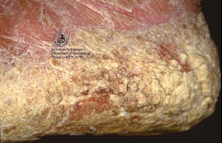
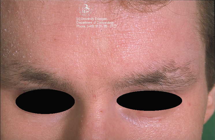
a. Papulosquamous disease(1) Seborrhea and dandruff:scalp, face, palmar,eyelid(2) Psoriasis: nails, nail pitting, nail oil spot change, knee, plantar, scalp, Plaque Type, Pustular Psoriasis, Guttate Type (1),(2), Auspitz Sign, heel, hypertrophic plaque, Psoriatic Arthritis of the hands
(3) Pityriasis rosea: close-up
(4) Superficial fungal infections
(a) Dermatophyte:Tinea Corporis, T.Faciea, T. Capitis(b) Candidiasis: Hairy Tongue, buccal mucosa, perleche, groin
(c) Tinea Versicolor
(5) Miliaria (prickly heat)
(6) Lichen Planus: buccal mucosa (1),(2), nail dystrophy, wrist
b. Vesiculobullous diseases
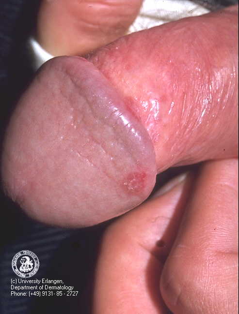
(1) Impetigo: paranasal(2) Herpes Simplex: recurrent labial ,penis
(3) Herpes Zoster (shingles), forehead
(4) Varicella
(5) Pemphigoid: abdomen, axillae
(6) Pemphigus: back, buccal mucosa, labial, vulgaris, drug induced
(7) Dyshidrosis pompholyx
(8) Erythema multiforme: palmar (1), dermatostomatitis
(9) Dermatitis herpetiformis, arm
(10) Epidermal necrolysis, (Stevens-Johnson Syndrome)
(11) Epidermolysis bullosa: (Dowling-Meara)
c. Dermatitis
(1) Contact: Contact Dermatitis Patterns finger, acute, vesicles and blisters, hands, chronic,wrist, foot (shoe), chemophotodermatitis,torso and arms,patch test,photodermatitis, neck , chemical burn(2) Atopic: chronic, antecubital fosse, chest, earlobe, face , finger, finger (2), hyperlinear palm , hyperlinear hands, labial, popliteal fossae, Dennie Morgan lines, Dirty Neck, Hertog's Sign (1), Hertog's Sign (2), Nipple Eczema , perleche, perleche (2),
(3) Generalized exfoliative: erythroderma
(4) Nummular: lower abdomen
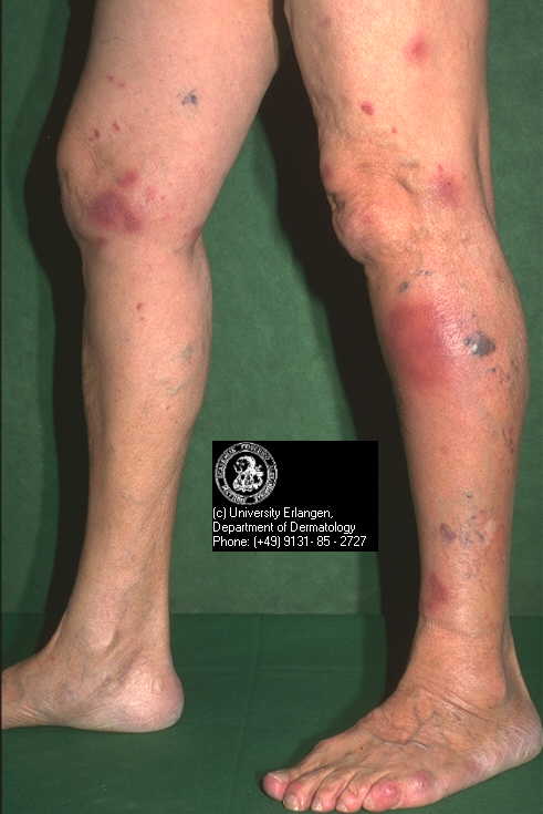
(5) Stasis (stasis ulcer):legs (1), (2)
(6) Diaper rash, candidal
d. Macular eruptions
(1) Viral exanthems: erythema infectiosum, Meningococcemia(2) Drug eruptions (Stevens-Johnson Syndrome)
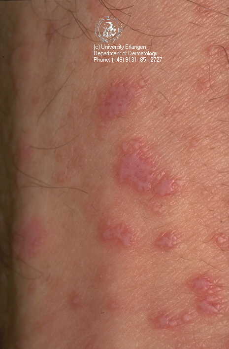
e. Urticarial eruptions
(1) Urticaria (hives) elbow,(2) Dermatographism: reverse dermatographism
f. Nodules
(1) Erythema nodosum:(1),(4)Sarcoid
(5) Deep fungal infections: Nocardia Braziliensis (1), (2) , Sporotrichosis (1), (2)
(6) Cysts:Eccrine spiradenoma
g. Other pruritic conditions
(1) Generalized(a) Scabies: diagram, scrotom(b) Dry skin (asteatosis)
(c) Secondary systemic disease
(2) Localized
(a) Lichen simplex chronicus (localized neurodermatitis)(b) Cutaneous papular amyloidosis
(c) Pruritis ani
(d) Pediculosis (lice)
(e) Chigger and other insect bites: Brown Recluse Spider Bite
h. Cutaneous infections
(1) Bacterial(a) Impetigo (see vesiculobullous disease): Paranasal , Bullous Impetigo, possible psoriatic diathesis(b) Erysipelas
(c) Lymphangiitis
(d) Cellulitis: Periorbital Cellulitis,Necrotizing Fasciitis
(e) Boil (e.g., furuncle, pustule, folliculitis, abscess, carbuncle, echthyma.) Eosinophilic Folliculitis:(1), (2), Gram Negative
(2) Mycobacterial
(a) Atypical mycobacteria
(3) Fungal
(a) Superficial fungal(b) Deep fungal
(4) Viral
(a) Herpes simplex: Herpetic whitlow (1), (2),(d) Molluscum contagiosum: penis
i. Complexion and cosmetic problems
(1) Acne vulgaris: (1), (2), (3)(3) Oily skin
(4) Enlarged pores
(5) Milia
(6) Vascular lesions: Varicose Veins, Tongue
(7) Wrinkles
j. Cutaneous injuries
(1) Burns(a) Thermal(b) Chemical
(c) Sunburn
(2) Blister
(3) Abrasion
(4) Laceration
(5) Bruise
(a) Trauma(b) Spontaneous purpura: Vasculitis
(6) Bites and stings: Rattlesnake Bite
k. Pigment disorders
(1) Hyperpigmentation(a) Generalized(b) Localized
i) Tinea versicolor (see superficial fungal infections)ii) Pityriasis alba: arm
iii) Vitiligo
l. New growths
(1) Benign(a) Inflammatory lesionsi) Acne cyst (see complexion problems) Inflammatory Acne, back, scarringii) Boil (see bacterial infection)
iii) Hidradenitis
iv) Pyogenic granuloma Granuloma faciale
(b) Hyperplasia
i) Verruca (common, plantar, anogenital, flat)iii) Corn and callus: Cutaneous Horn
iv) Epidermal cyst
v) Skin tag (acrochordon)
vi) Xanthelasma
(c) Neoplasia
i) Seborrheic keratosis: Seborrheic Keratosisii) Mole, nevus (intradermal, junctional, compound, halo, blue, congenital)
iii) Lipoma
v) Keloid: right lateral thigh
vi) Hemangioma: wrist
vii) Neurofibroma
viii) Other, such as fibroma, leiomyoma
(2) Premalignant
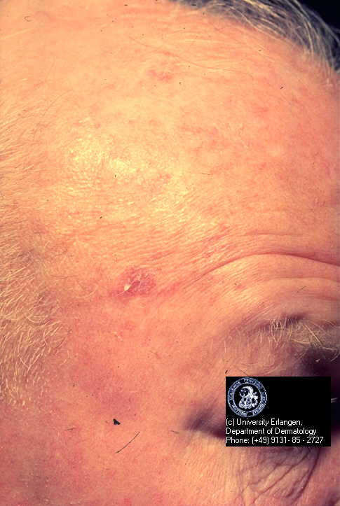
(a) Squamous cell carcinoma in situ (Bowen's Disease):dorsal arm, penis(b) Actinic Keratosis: (1),(2),(3)
(c) Actinic cheilitis: Actinic Cheilitis , Actinic Cheilitis, Lower Lip
(d)Disseminated superficial actinic porokeratosis
(e) Leukoplakia: tongue , Leucoplakia, Buccal Mucosa
(f)Keratoacanthoma, clear cell acanthoma
(g) Erythroplakia Erythroleucoplakia
(h) Premelanoma
(ii) Giant congenital nevus(iii) Dysplastic Nevus Syndrome
(iv) Radiation effects
(3) Malignant
(a) Basal cell carcinoma: small tumor (1) , large tumor, right cheek (2),(b) Squamous cell carcinoma: tongue, Lower Lip (1) ,(2) , floor of the mouth (1)
(i) Major clinical categories(ii) Prognostic and therapeutic guidelines
(d) Paget's disease (1)
(e) Cutaneous lymphoma
(f) Kaposi's lymphoma: (1) ,(2)
(g) Metastases to the skin, SCC
m. Cutaneous manifestations of systemic disease, including human immunodeficiency virus infection: Apthosis , Candidiasis, Kaposi's sarcomaoral, foot,n. Occupational skin disease
2. Hair problems
a. Fungal infection
b. Nonscarring alopecia
(1) Androgenetic alopecia (male pattern): Alopecia Universalis(2) Alopecia (areata (1), (2) /totalis/universalis): Trachonychia, Nail Pitting
(3) Telogen effluvium
(4) Traction and trichotilomania
(5) Endocrine effects
c. Scarring alopecia
(1) Discoid lupus (1 )(2) Lichen planopilaris
d. Ingrown hair (pseudofolliculitis)
e. Hypertrichosis
(1) Localized(2) Virulizing causes of hypertrichosis (generalized)
f. Texture alterations (hair dystrophy)
3. Nail problems
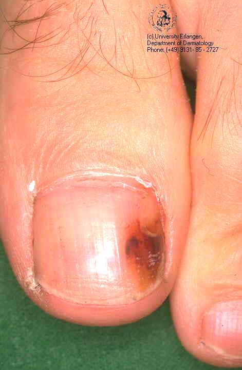
a. Trauma: Nail Tic Habit , Traumatic Nail remnantb. Disturbances associated with other dermatoses Splinter Hemorrhages, Nails
c. Disturbances associated with systemic disease
d. Texture alterations
(1) Brittle, weak nails(2) Thyroid disease
(3) Vascular insufficiency
(4) Lichen planus: Twenty Nail Dystrophy
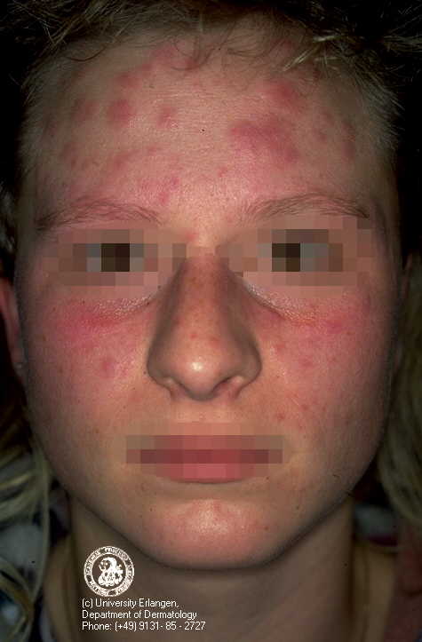
(5) Autoimmune disease changes (lupus erythematosus, scleroderma, etc.)
e. Fungal infection: Onychomycosis (1), (2), Chronic Mucocutaneous Candidiasis
f. Periungual and subungual conditions
(1) Ingrown nail(2) Paronychia
(3) Hematoma Resolving Subungual Hematoma
g. Colored nails: Melanonychia
h. New growths
(1) Benign
(a) Inflammatoryi) Granulomaii) Warts
(b) Neoplasia
(2) Malignant
(a) Melanoma(b) Squamous Cell Carcinoma
4. Mucous membrane lesions
5. Oral lesions
a. Thrushb. Mouth ulcers
c. Sicca
e. Geographic tongue
f. Black hairy tongue Brown Hairy Tongue
g. Leukoplakia
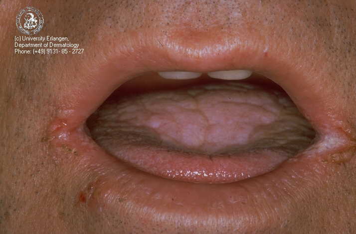
h. Cheilitis Perleche, Candida, angular cheilitis
Skills
A. Diagnostic skills
1. Performance of history and physical with differential diagnosis
2. Acquisition, examination and interpretation of laboratory specimens
a. Biopsyb. Culture
c. Scraping
3. Skin testing techniques and interpretation
4. Use of mechnaical devices, i.e., Wood's light
5. Systematic evaluation (if indicated)
6. Description of distribution and character of lesions
B. Management skills
1. Genetic counseling
2. Nutrition counseling
3. Peventive care
a. Routine skin careb. Avoidance of environmental causes
c. Sunscreens
d. Appropriate use of over-the-counter lotions
4. Health promotion
5. Patient education
6. Use of photographs to document progress
7. Use of scales/indices to grade disease severity
8. Use of consultations and referrals
C. Therapeutic skills
1. Medical
a. Topicalb. Systemic
2. Surgical
a. Cauterization of skin lesions(1) Acid cautery(2) Electrocautery
(3) Electrodessication and curettage
b. Cryosurgeryc. Punch biopsy
d. Excision of skin lesions
e. Intralesional injection of corticosteroids
f. Incision and drainage
g. Treatment of ingrown toenails