
COMMON DERMATOLOGIC DISEASES
Rhett J. Drugge, MD
COMMON DERMATOLOGIC DISEASES
Rhett J. Drugge, MD
Tinea versicolor is caused by the Malassezia furfur. It seems to be increasingly common, especially in people who habitually lie on sandy beaches in the Caribbean. It usually affects the upper trunk and produces a variegated hyper-or hypopigmentation. Selenium sulfide shampoo, using 2% selenium sulfide two or three time a week and leaving it on overnight, is very helpful; and if the areas are localized, Whitfield's ointment, 3% sulfur-sal and Whitfield's"ointment" in an alcoholic solution are effective preparations. Twenty percent or a saturated solution of sodium hyposulfite (or sodium thiosulfate) is a classical remedy, but has an unpleasant odor. When people perspire in hot communities, it is unpleasant for their friends.
Ketoconazole 200 mg per day for two days in an adult is effective. Efficacy should be improved by ingestion with a carbonated beverage, followed by engaging in a sweat producing activity without an ensuing shower. This approach will leave a residue in the sweat. Caution should be taken as ketoconazole has a potent inhibitory effect on microsomal P-450 hydroxylation, thereby producing derangements in the metabolism of lipophilic drugs and endogenous molecules. The short duration of treatment makes the fatal hepatic necrosis occasionally seen with this drug of little or no consequence.
The problem of cure is largely due to the fact that the small
fungus can get down into the hair follicles, and when a clinical cure
appears to have been achieved, the infection may still be present in
the depths of the hair follicles. Then, when the patient perspires
excessively, the organism comes back up onto the surface again and
reinfects, and the clinical infection is obvious. The patient
therefore may need to continue to treat himself intermittently for
years with anti-dandruff shampoos containing mild antimycotic agents
such as selenium sulfide. ![]()
This disease, caused by the Cladosporium werneckii or
mansonii (these are synonyms for Hormodendrum), occurs
in Brazil, Cuba, Panama, Puerto Rico and India. In the Western
hemisphere, it involves only the palms. There are brown or black
asymptomatic annular lesions, which usually respond to keratolytics,
e.g., 5% sulfa-sal. ![]()
Trichosporosis causes nodes on the hair which are white, brown or black. There are essentially two varieties: white and black. It occurs in patients in the tropics, with occasional cases in the temperate zones, mostly on the scalp. It does not fluoresce under Wood's light. Usually the treatment is shaving, or formalin preparations.
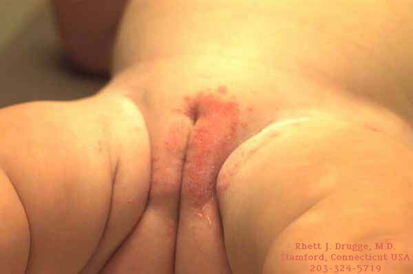
Small yellow-red or black axillary spots form and treatment is
with an alcoholic solution of formaldehyde 2%, or a sulfur 5%
ointment. It fluoresces under Wood's light, and clothing is usually
stained by the colored sweat. Oral antibiotics and antibiotic
vanishing creams are a somewhat more elegant way to clear these
patients. ![]()
Erythrasma is caused by a gram-positive bacteria, Corynebacterium
minutissimum, occurring in humid climates. It fluoresces with a coral
red color and responds to systemic antibiotics, especially
erythromycin, tetracycline and chloromycetin; and occasionally in
widespread cases, this is the only way you can cure them. Penicillin
and griseofulvin do not help, and generally keratolytic ointments are
sufficient to clear them. ![]()
Ringworm fungi consist of Microsporum, Epidermophyton and
Trichophyton. Human serum inhibits the growth; this is diminished in
blood dyscrasia and possibly also in schizophrenia. There is evidence
to suggest that the infections are self-limiting, and in individuals
who lack this "serum factor," the mycelia proliferate in the dermis
in the living tissue and not just in the keratogenous zone, which is
a nonliving keratinized structure. Individuals may get granulomas;
e.g., women who shave their legs may get a hair follicle granuloma
(Majocchi's Granuloma). ![]()
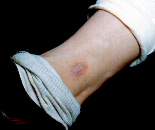
Tinea capitis is caused by any dermatophyte except Epidermophyton floccosum and Trichophyton concentricum. Microsporum usually fluoresce with a bright green color, chiefly M. canis, and M. audouinii. These usually clear at puberty, except for a small percentage of favus and endothrix, Trichophyton infections, e.g., the tonsurans and violaceum infections. Chronicity is associated with a lack of inflammatory response, and while kerion is curative in many cases, it is extremely unpleasant.
T. tonsurans is the currently endemic in the United States and is the chief cause there of tinea capitis. T. violaceum and M. gypseum, contracted from soil and animals, do not fluoresce and can affect adults. Most other microsporum affect children, but not adults. Occasionally in the tropics, T. rubrum can produce tinea capitis, and then it is endothrix. Cultures should always be made to identify the fungus. Rapid diagnosis can be made by placing epilated hair on a glass slide, applying a few drops of 10% KOH, placing a coverslip on top, heating over an alcohol lamp and examining with a low-power microscope directly.
Griseofulvin is curative for most cases of tinea capitis. For persons weighing less than 30 lb., the dose of ultramicrosized griseofulvin is 0.25 gm per day; 50 lb. and over, 0.375 gm per day; and 100 lb. and over, 0.5 gm a day.
Many people with cases of tinea tonsurans have to be kept on this
treatment for 3 or 4 months. ![]()
This is often mistaken for a bacterial folliculitis at first.
M. canis has a positive reaction to Wood's light but other
fungi do not and have to be identified by cultures of epilated hair.
Generally T. mentagrophytes produces dry inflammatory nodules,
while T. rubrum and others produce more chronic infections
similar to sycosis barbae. Treatment with griseofulvin for several
months clears almost all cases. ![]()
Annular or polycyclic marginated scaly erythemas, occasionally
plaques suggesting psoriasis
, and less often granulomatous nodules are found. Verrucous lesions
are rare. ![]()
This disorder is generally candidiasis
if the scrotum is involved, otherwise the classic jock itch with the
distinct margin can be caused by E. floccosum, T.
rubrum or T. mentagrophytes. ![]()
Tinea is vesicular if caused by T. mentagrophytes and dry,
branny and scaly if caused by T. rubrum. Occasionally monilia
will produce a margin halfway up the palm. The most common pattern of
hand involvement is for a single hand to be involved while both feet
are involved in a moccasin distribution scaling typical of T.
rubrum. ![]()
Local treatment of dermatophytes makes up probably 5% of a physician's practice. Griseofulvin has certainly helped in the management of these cases, particularly the T. rubrum cases, but it is still amazing how many people do not seem to get complete clearing with oral griseofulvin. Micronizing the powdered tablet or capsule and administering this along with a fatty meal greatly assist in absorption. The side effects of occasional headache and occasional depression and nausea can generally be dealt with, but local treatment is still the crux of the management of most dermatophytes, particularly athlete's foot. Itraconazole, at an adult dose of 200 mg per day is an effective alternative therapy in resistant cases. Undecyclenic acid, 20% powder or cream, is effective. Propionic acid and tolnaftate or Tinactin (Schering), either cream or solution, are effective if rubbed on a dry area twice a day at least and rubbed in thoroughly. Halotex and Micatin cream and Lotrimin cream are effective remedies.
Whitfield's ointment, which is 6% salicylic acid and 12% benzoic
acid in a suitable base, usually lanolin or Vaseline, though we can
cut the strength and use it in USP hydrophilic ointment, is very
effective in treating the blistering type of dermatophytosis. Three
percent sulfur, 3% salicylic acid in a suitable base, and
occasionally anywhere from 2% to 5% ammoniated mercury in a suitable
base are also suitable although more confined to the more chronic
types of dermatophytosis. In acute blistering types of T.
mentagrophytes athlete's foot, soaks and Burrow's solution or
potassium permanganate, or some astringent type of wet dressing that
shrivels the blisters and helps to pass the disorder from the acute
stage to the more amenable-to-therapy second stage, is necessary.
Very often steroids will assist this. ![]()
Acute
Contact Dermatis ,subacute, chronic
Atopic
eczema
Venous
Dermatitis, Venous Eczema, Stasis Dermatitis
Palmar
Eczema
Tinea
Nummular
eczema
Lichen simplex chronicus
![]()
Impetigo
contagiosa- staph, strep
Erysipelas
bullous impetigo - staph
Primary
Cutaneous Tuberculosis
![]()
A formerly fatal bullous disease shows acantholysis as a constant feature. The separation is chiefly above the basal layer in the epidermis. Nikolsky's sign, or the ability to extend the margin of the bulla peripherally by pressure, is a diagnostic feature. The etiology is obscure, although a degeneration of the epidermal cell with loss of the intercellular bridges is an early feature.
Lesion skin shows direct and indirect immunofluorescent studies with intraepidermal intercellular deposition of IgG and C3 (complement) consistently. Titers on indirect serum immunofluorescence (IF)reflect the activity of the disease.
Oral lesions are almost always present, sometimes extensively, and the disease may start in the oropharynx. Bullous lesions occur in crops on normal skin with no itching, no erythema surrounding(as with erythema multiforme) and no grouping, and circinate forms on the shoulders, buttocks and axilla, (Fig. 6-1), as with dermatitis herpetiformis (which itches). The bullae are flaccid and rupture easily, leaving a raw surface.
Systemic steroids are the method of choice for initial treatment.
The dose must usually be high at first, then gradually stepping down
(start with 60-150 mg per day of prednisone). If the process is not
controlled in a week, the dose should be doubled and then
stepped down gradually. Antibiotic support with tetracycline...
![]()
Psoriasis
Seborrheic
dermatitis
Pityriasis
rosea
Secondary syphilis
Lichen
planus
Tinea
Parapsoriasis - mycosis fungoides
Drug reactions (Cutaneous
Drug Reactions Database )
Erythema
Chronicum Migrans
![]()